Sexual activity has been the subject of fascination for humans since the dawn of civilization. Research shows that sexual activity can have a significant impact on the brain and the body.
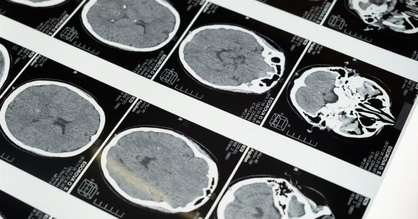
MRI scans now offer scientists a fascinating tool to study how different parts of the brain are activated during sexual activity.
The brain and sexual arousal
Sexual arousal is a complex process that involves several different areas of the brain. The brain’s limbic system, which is responsible for emotions and motivation, plays a crucial role in sexual arousal.
The amygdala, which is located in the temporal lobe, is believed to be responsible for triggering sexual attraction in response to visual or physical stimuli. The hypothalamus, which is located in the center of the brain, controls various physiological responses to sexual stimuli, such as increasing blood flow to the genitals and increasing heart rate.
The impact of sexual activity on the brain
A study published in 2017 by the Max Planck Institute for Human Development in Berlin used functional magnetic resonance imaging (fMRI) to study the brain activity of 14 women while they were sexually aroused.
The study found that sexual arousal activates a number of different areas of the brain, including the amygdala, the thalamus, and the insula. The insula is located in the cerebral cortex and is responsible for processing information related to physical sensations and emotions.
The study also found that sexual arousal decreases activity in the prefrontal cortex, which is responsible for decision-making, planning, and judgment.
This reduction in activity in the prefrontal cortex may help explain why people sometimes make decisions while sexually aroused that they later regret.
Different types of sexual activity and brain activity
A study published in 2015 by the University of Groningen in the Netherlands used fMRI scans to study brain activity while participants were either masturbating or having sex with a partner.
The study found that different parts of the brain were activated depending on the type of sexual activity.
When participants were masturbating, the study found that activity in the amygdala, the hippocampus, and the anterior cingulate cortex was significantly lower than when they were having sex with a partner.
The anterior cingulate cortex is located in the frontal part of the brain and is associated with pain and emotion. The hippocampus is located in the temporal lobe and is responsible for processing memories.
The study also found that activity in the striatum, which is responsible for motivation and reward, was higher during sexual activity with a partner than during masturbation.
The striatum is located in the center of the brain and plays an essential role in the reward system.
The impact of orgasm on the brain
Orgasm is a complex physiological process that involves the activation of several different areas of the brain. During orgasm, the brain releases a flood of neurotransmitters and hormones, such as dopamine, serotonin, and oxytocin.
A 2017 study from Rutgers University used fMRI scans to study brain activity during orgasm. The study found that orgasm activates several different areas of the brain, including the prefrontal cortex, the temporal lobe, and the cerebellum.
The prefrontal cortex is responsible for decision-making and behavior. The temporal lobe plays a crucial role in memory and emotion. The cerebellum is responsible for movement control and coordination.
The study also found that activity in the amygdala, the hypothalamus, and the ventral striatum was significantly decreased during orgasm. The ventral striatum is an essential part of the reward system and is associated with pleasure and motivation.
The study suggests that decreased activity in these areas of the brain may help explain the intense pleasure and relaxation that people experience during orgasm.
The impact of sexual desire on the brain
Sexual desire is a complex phenomenon that involves the interaction of various psychological, emotional, and physical factors. Research suggests that sexual desire can have a significant impact on the brain.
A study published in 2011 in the Journal of Sexual Medicine used fMRI scans to study brain activity in men and women while they were sexually aroused.
The study found that sexual desire is associated with increased activity in the ventromedial prefrontal cortex, which is responsible for processing emotions and decision-making.
The study also found that sexual desire is associated with decreased activity in the amygdala, which is responsible for processing emotions, especially fear and anxiety.
This finding suggests that sexual desire may help reduce anxiety and stress levels.
The impact of sexual orientation on the brain
Sexual orientation is a complex phenomenon that has yet to be fully understood by scientists. Research shows that sexual orientation can have a significant impact on the brain.
A study published in 2017 by the University of Montreal used fMRI scans to study brain activity in heterosexual and homosexual men while they were sexually aroused.
The study found that sexual orientation is associated with differences in brain activity in several areas of the brain, including the hypothalamus and the amygdala.
The study found that heterosexual men had higher activity in the amygdala than homosexual men, suggesting that heterosexual men may be more sensitive to visual sexual stimuli.
The study also found that homosexual men had higher activity in the hypothalamus than heterosexual men, suggesting that sexual orientation may be related to differences in the response of the hypothalamus to sexual stimuli.
Conclusion
The study of the brain and sexual activity is a rapidly evolving field that offers fascinating insights into human behavior and physiology.
MRI scans provide researchers with a powerful tool to study the brain and the impact of sexual activity on different brain regions. While much remains to be discovered, these studies offer exciting possibilities for future research and understanding of human sexuality.


